The female pelvis is a compact anatomical neighborhood where the urinary, reproductive, digestive, muscular, nervous, and connective-tissue systems share very limited real estate. Somehow, the bladder, uterus, vagina, ovaries, bowel, nerves, blood vessels, and multiple layers of muscle cooperate without holding weekly planning meetings.
Understanding a female pelvis diagram becomes much easier when you stop viewing it as a collection of mysterious labels. Each structure has a position, a job, and important relationships with the organs around it. This guide explains those relationships, identifies the major inner pelvic muscles, and shows how the entire region supports urination, bowel movements, sexual function, pregnancy, childbirth, posture, and movement.
What Does the Female Pelvis Contain?
The pelvis is the basin-shaped bony region below the abdomen. Its framework includes the two hip bones, sacrum, coccyx, pubic symphysis, and several joints. Inside this framework sit organs belonging to three major systems:
- Urinary system: the bladder, urethra, and lower portions of the ureters.
- Reproductive system: the ovaries, uterine tubes, uterus, cervix, and vagina.
- Digestive system: portions of the small and large intestines, sigmoid colon, rectum, and anal canal.
The pelvic floor forms the muscular base beneath these organs. Ligaments and fascia provide additional support from the sides and above. Anatomy varies naturally, and surgery, pregnancy, age, congenital differences, or medical conditions may change which structures are present and where they sit.
A Simplified Side-View Female Pelvis Diagram
In a typical side-view diagram, the bladder is toward the front, the uterus and vagina occupy the middle, and the rectum lies behind them. This arrangement explains why a condition affecting one organ can produce symptoms elsewhere. A full rectum may increase bladder pressure, for example, while a prolapsing bladder can press into the front wall of the vagina.
Female Pelvic Organs and Their Functions
Bladder and Urethra
The bladder is a hollow muscular organ located behind the pubic bone and in front of the uterus and vagina. Its flexible walls expand as urine arrives through the ureters. During urination, the bladder muscle contracts while the urethral sphincters and pelvic floor relax in a coordinated sequence.
The urethra is the short tube that carries urine from the bladder to the outside of the body. The pelvic floor helps support the bladder neck and urethra. When pressure rises during a cough, jump, sneeze, or enthusiastic laugh, healthy support tissues help keep the urethra closed. If that system becomes weak or poorly coordinated, stress urinary leakage may occur.
Ovaries
The ovaries are paired reproductive organs usually positioned on either side of the uterus. They contain follicles that may release an egg during ovulation and produce hormones, including estrogen and progesterone. These hormones influence the menstrual cycle, reproductive tissues, bones, cardiovascular health, and many other body processes.
Although diagrams often show the ovaries sitting symmetrically like two perfectly placed decorative lamps, their exact positions can vary. They are mobile structures held by ligaments and surrounded by blood vessels, nerves, and nearby bowel.
Uterine or Fallopian Tubes
The uterine tubes extend from the upper uterus toward the ovaries. Their fingerlike ends, called fimbriae, help guide an ovulated egg into the tube. Fertilization most commonly occurs within a uterine tube, after which the developing embryo normally travels toward the uterine cavity.
The tubes are not sealed directly onto the ovaries. Instead, each tube opens into the pelvic cavity near an ovary. That detail surprises many people and explains why pelvic infections or conditions such as endometriosis can affect multiple nearby structures.
Uterus and Endometrium
The uterus is a hollow, muscular organ commonly described as pear-shaped. It usually sits between the bladder and rectum, although its angle varies. Some uteruses tilt forward, while others tilt backward; either position can be a normal anatomical variation.
The thick muscular wall, called the myometrium, contracts during menstruation and labor. The inner lining is the endometrium. During a menstrual cycle, this lining changes in response to hormones. If pregnancy does not occur, much of it is shed during menstruation. If implantation occurs, the uterus expands dramatically to support the developing pregnancy. Few organs can remodel themselves so impressively without demanding a home-renovation permit.
Cervix
The cervix is the lower, narrower portion of the uterus. It projects into the upper vagina and contains a central cervical canal. Menstrual fluid passes through this canal, sperm may travel through it, and cervical mucus changes during the menstrual cycle.
During pregnancy, the cervix normally remains closed and helps support the pregnancy. During labor, it softens, thins, and dilates. Because cervical cells can undergo precancerous changes, routine cervical cancer screening may be recommended according to a person’s age, health history, and current medical guidelines.
Vagina
The vagina is an elastic muscular canal extending from the cervix to the vaginal opening. It permits menstrual flow to leave the body, receives penetration, and forms part of the birth canal. The vagina is internal; the vulva refers to the external genital structures.
The front vaginal wall is closely related to the bladder and urethra, while the back wall lies near the rectum. The upper vagina is supported by connective tissue and ligaments. These relationships are clinically important because weakened support may allow the bladder, rectum, uterus, or vaginal apex to bulge toward the vaginal canal.
Rectum and Anal Canal
The rectum is the final storage region of the large intestine. When stool enters it, nerves signal the need for a bowel movement. Emptying requires the rectum, anal sphincters, abdominal wall, and pelvic floor to coordinate correctly.
In particular, the puborectalis muscle must lengthen and relax so the anorectal angle can open. If the pelvic floor tightens instead of relaxing, a person may strain, feel incompletely emptied, or experience constipation despite having no physical blockage.
Inner Female Pelvic Muscles
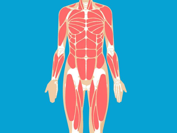
The phrase “inner pelvic muscles” may refer to the pelvic floor itself, the muscles lining the pelvic walls, or both. They work with the diaphragm, abdominal muscles, back muscles, hips, and connective tissues as part of an integrated pressure-management system.
Levator Ani
The levator ani forms most of the pelvic floor. It is generally divided into three overlapping components:
- Puborectalis: forms a sling around the rectum and helps maintain the anorectal angle, supporting bowel continence.
- Pubococcygeus: stretches from the pubic region toward the coccyx and nearby tissues. It supports pelvic organs and contributes to urinary, bowel, and sexual function.
- Iliococcygeus: forms a thinner but broad supportive layer extending toward the coccyx and central connective tissues.
Together, these muscles lift and support the pelvic contents. They also contain openings for the urethra, vagina, and anal canal. The floor is therefore less like a solid wooden platform and more like a responsive trampoline with carefully controlled passageways.
Coccygeus
The coccygeus, also called the ischiococcygeus, lies toward the back of the pelvic floor. It extends between the ischial spine and the lower sacrum and coccyx. It reinforces the posterior pelvic floor and may help stabilize or gently draw the coccyx forward after it moves during bowel movements or childbirth.
Obturator Internus
The obturator internus covers much of each side wall of the pelvis. Its tendon leaves the pelvis and attaches to the femur, allowing the muscle to rotate and stabilize the hip. Pelvic floor connective tissue attaches along fascia covering the obturator internus, creating a direct functional relationship between hip mechanics and pelvic support.
This connection is one reason pelvic floor rehabilitation may include hip exercises rather than an endless parade of Kegels.
Piriformis
The piriformis lies along the back wall of the pelvis and travels through the greater sciatic foramen to the femur. It helps rotate and stabilize the hip. The sciatic nerve usually passes beneath it, while important pelvic nerves and blood vessels travel nearby. Piriformis tension can contribute to buttock or hip symptoms, although not every ache in the neighborhood should automatically be blamed on this muscle.
Deep Perineal and Sphincter Muscles
Smaller muscles around the urethra, vagina, and anus contribute to continence, sexual function, and stabilization of the openings through the pelvic floor. These include components of the external urethral sphincter complex, external anal sphincter, deep transverse perineal muscles, bulbospongiosus, and ischiocavernosus.
They do not work in isolation. Their performance depends on sensation, nerve signaling, connective-tissue support, breathing, and the ability to contract and release at the correct time.
How the Pelvic Floor Functions
Organ Support and Pressure Control
The pelvic floor supports the bladder, uterus, vagina, and bowel while responding to pressure from above. When a person coughs or lifts something, the diaphragm and abdominal wall change pressure inside the trunk. The pelvic floor should respond automatically, absorbing and managing that force without allowing leakage or excessive organ movement.
Bladder and Bowel Control
Pelvic muscles contract to help delay urination, gas, or a bowel movement. They must then relax so the bladder and bowel can empty efficiently. Strength matters, but timing and relaxation matter just as much. A muscle that remains clenched all day is not necessarily strong; it may simply be overworked and tired.
Sexual Function
The pelvic floor contributes to genital sensation, arousal, vaginal tone, and rhythmic contractions during orgasm. Excessive tension may cause pain with penetration, difficulty using tampons, or discomfort during pelvic examinations. Weakness may reduce sensation or contribute to pelvic heaviness, but sexual symptoms can have hormonal, neurological, emotional, vascular, and relationship-related causes as well.
Pregnancy and Childbirth
During pregnancy, the pelvic floor supports increasing weight while adapting to hormonal and postural changes. During vaginal birth, its tissues must stretch considerably. Recovery differs from person to person and may involve weakness, muscle injury, scar sensitivity, prolapse symptoms, or excessive protective tightening.
Core Stability and Movement
The pelvic floor works with the diaphragm, deep abdominal muscles, spinal stabilizers, and hip muscles. It subtly responds while walking, lifting, breathing, and changing position. Good pelvic health is therefore not about squeezing one hidden muscle as hard as possible. It is about coordination across the entire trunk and pelvis.
When Pelvic Muscles Become Weak or Too Tight
Signs of Weakness or Reduced Support
- Urine leakage with coughing, running, jumping, or lifting
- Difficulty controlling gas or stool
- Pelvic pressure, heaviness, or a vaginal bulge
- Reduced support during physical activity
- A feeling that the bladder or bowel does not empty normally
Pregnancy, childbirth, aging, menopause, pelvic surgery, chronic coughing, constipation, repeated straining, obesity, and frequent heavy lifting may increase pressure on pelvic support tissues. Genetics and connective-tissue characteristics also influence risk.
Signs of Excessive Tension
- Pelvic, vaginal, rectal, tailbone, or lower abdominal pain
- Pain with penetration or a pelvic examination
- Urinary urgency, frequency, or difficulty starting the stream
- Constipation or straining despite soft stool
- A persistent feeling that the pelvic muscles cannot let go
For a tight or poorly relaxing pelvic floor, more strengthening may worsen symptoms. Treatment may instead emphasize breathing, relaxation, mobility, bowel mechanics, manual therapy, and gradual retraining under the guidance of a pelvic floor physical therapist.
Practical Ways to Support Pelvic Health
Pelvic floor exercises can help some people, particularly when weakness contributes to urinary leakage. A correct contraction should create a gentle inward-and-upward lift around the urethral, vaginal, and anal openings. The buttocks, thighs, and abdomen should not perform the entire exercise while the actual pelvic floor takes a coffee break.
Avoid repeatedly stopping urine midstream as a workout. That maneuver may help identify the muscles once, but routinely interrupting urination can interfere with normal emptying. It is also important to release fully between contractions and continue breathing.
Other helpful habits may include treating constipation, avoiding habitual straining, gradually managing heavy loads, maintaining comfortable physical activity, addressing chronic coughing, and allowing enough time for postpartum recovery. Symptoms that persist deserve an individualized assessment rather than a generic exercise sheet.
Real-World Experiences: What Pelvic Function Can Feel Like
The following illustrative experiences combine patterns commonly encountered in pelvic health care. They are not personal medical stories and cannot diagnose an individual condition.
Experience 1: Leakage After Childbirth
Imagine a new parent who feels mostly recovered several months after delivery but leaks a small amount of urine whenever she sneezes or jogs. She assumes this is simply the permanent membership fee for having a baby. During an evaluation, however, a pelvic floor therapist finds that her muscles can contract but do not respond quickly when abdominal pressure rises.
Her rehabilitation focuses on gentle contractions, complete relaxation, breathing, hip strength, and practicing pelvic floor activation before a cough or lift. Over time, she returns to running gradually. The important lesson is that common postpartum symptoms are not necessarily symptoms a person must quietly accept forever.
Experience 2: When More Kegels Are the Wrong Answer
Another person experiences urinary urgency, constipation, and pain with penetration. She begins performing dozens of strong Kegels every day because the internet has apparently appointed Kegels manager of every pelvic problem. Her discomfort increases.
An examination reveals that her pelvic floor is already overactive and has difficulty lengthening. Treatment emphasizes diaphragmatic breathing, relaxation, comfortable hip mobility, bowel-position adjustments, and learning to release the pelvic muscles. Her experience shows why “weak” and “tight” are not interchangeableand why symptoms alone may not reveal which pattern is present.
Experience 3: Pelvic Heaviness During Exercise
A recreational weightlifter notices vaginal heaviness after high-intensity sessions. She does not have sharp pain, but the sensation feels as though something is pressing downward. Instead of abandoning exercise, she seeks an evaluation. The clinician identifies mild pelvic organ prolapse and reviews breathing, load management, constipation prevention, and exercise modifications.
With individualized guidance, she continues strength training while monitoring symptoms. Some people with prolapse remain highly active; the best plan depends on symptom severity, anatomy, goals, and the body’s response to different movements.
Experience 4: Changes Around Menopause
A woman around menopause develops vaginal dryness, urinary urgency, and discomfort that she initially blames entirely on aging. Her clinician explains that declining estrogen can affect vaginal and urinary tissues, while pelvic muscle tension may develop as the body braces against discomfort.
Her care combines appropriate medical treatment for tissue changes with pelvic floor therapy. Addressing both the organs and muscles works better than treating either one as an isolated culprit. Pelvic anatomy may be crowded, but its problems rarely respect departmental boundaries.
Experience 5: The Value of an Accurate Diagram
A patient with pelvic pain studies a female pelvis diagram before her appointment. Seeing the bladder in front of the vagina and the rectum behind it helps her describe where she feels pressure. She also realizes that “ovary pain” cannot be confirmed by location alone because muscles, bowel, bladder, nerves, and reproductive organs can produce overlapping sensations.
During the visit, she gives a clearer history: when the pain occurs, whether it changes with urination or bowel movements, and how it relates to menstruation and activity. The diagram does not diagnose her, but it gives her a useful vocabulary. That is the real value of anatomy educationnot encouraging people to play detective with every twinge, but helping them communicate accurately and participate in care.
When to Seek Medical Care
Schedule an evaluation for persistent pelvic pain, new urinary or bowel problems, pain during sex, unexplained bleeding, a vaginal bulge, or symptoms that interfere with daily life. Depending on the concern, care may involve a primary care clinician, gynecologist, urogynecologist, urologist, colorectal specialist, or pelvic floor physical therapist.
Seek urgent medical care for severe or rapidly worsening pelvic pain, especially when accompanied by pregnancy, fainting, dizziness, fever, vomiting, heavy bleeding, abdominal rigidity, or an inability to urinate or pass stool. Sudden pelvic pain can occasionally signal conditions such as ectopic pregnancy, ovarian torsion, a ruptured cyst, infection, or another emergency.
Conclusion
The female pelvis contains far more than reproductive organs. The bladder and urethra occupy the front, the uterus and vagina usually sit centrally, and the rectum lies behind them. Beneath this crowded arrangement, the levator ani, coccygeus, sphincters, and other inner muscles provide support while coordinating urination, bowel movements, sexual activity, breathing, movement, pregnancy, and childbirth.
Healthy pelvic muscles must be able to contract, relax, stretch, and respond at the right moment. Weakness can cause leakage or prolapse symptoms, while excessive tension can contribute to pain and emptying difficulties. A diagram offers an excellent map, but persistent symptoms require professional assessment because neighboring pelvic structures have a remarkable talent for impersonating one another.