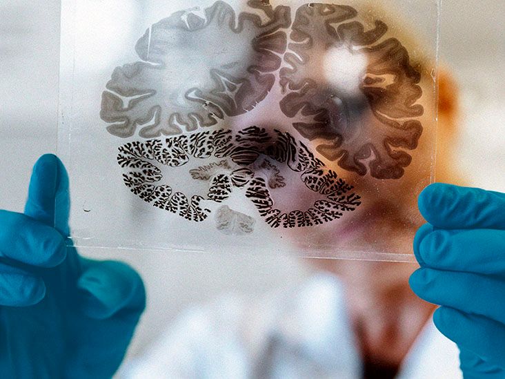
“Brain atrophy” is the kind of phrase that can make an otherwise calm person stare at an MRI report as though it has just delivered a personal threat. It sounds dramatic, but the term itself does not identify one particular disease. Brain atrophy, also called cerebral atrophy, describes a loss of brain cells and the connections between them, resulting in reduced brain volume.
A certain amount of brain volume loss can occur as part of normal aging. However, shrinkage that is unusually rapid, severe, or concentrated in specific regions may be associated with neurological disease, stroke, infection, injury, or another medical condition. The symptoms and outlook depend less on the word “atrophy” and more on what caused it, which areas are affected, and whether the process is stable or continuing.
What Is Brain Atrophy?
The brain contains billions of neurons that communicate through vast networks of connections. When neurons die or their connections deteriorate, the affected brain tissue may lose volume. Imaging studies may describe this change as brain volume loss, cerebral atrophy, cortical thinning, or regional atrophy.
Brain atrophy can be classified in two broad ways:
- Generalized or diffuse atrophy affects multiple areas throughout the brain.
- Focal or regional atrophy is concentrated in a particular structure or lobe.
The distinction matters because individual brain regions have different jobs. Shrinkage in memory-related structures may produce a different pattern of symptoms from shrinkage in areas controlling balance, vision, speech, judgment, or personality. In other words, the brain is not one giant on-off switch. It is more like an extremely complicated office in which every department insists it is essentialand, annoyingly, most of them are.
Does Brain Atrophy Always Mean Dementia?
No. Dementia is a clinical syndrome involving cognitive decline severe enough to interfere with daily life. Brain atrophy is a structural finding. A person may have mild volume loss without dementia, while another person may have substantial cognitive symptoms related to a particular pattern of regional damage.
Atrophy can support the diagnosis of Alzheimer’s disease or another form of dementia, but an MRI scan alone generally cannot establish the complete diagnosis. Clinicians must interpret imaging alongside symptoms, medical history, neurological findings, cognitive testing, laboratory results, and sometimes additional biomarkers.
Symptoms of Brain Atrophy
Brain atrophy does not have one universal symptom list. Some people have noticeable difficulties, while others have mild imaging changes discovered during a scan performed for an unrelated reason. Symptoms depend on the amount, location, cause, and speed of tissue loss.
Cognitive Symptoms
When atrophy affects networks involved in thinking and memory, possible symptoms include:
- Increasing forgetfulness
- Difficulty learning new information
- Reduced attention or mental processing speed
- Trouble planning, organizing, or solving problems
- Confusion about dates, places, or familiar routines
- Poor judgment or difficulty managing finances
- Problems completing multistep activities
Occasionally forgetting where the keys are is common. Finding them in the freezer repeatedlyand being unable to explain how they got theredeserves more attention.
Language and Communication Problems
Damage involving language networks may cause difficulty finding words, naming familiar objects, understanding speech, reading, writing, or forming clear sentences. In frontotemporal disorders, language problems may be among the earliest symptoms rather than appearing late in the illness.
Personality, Mood, and Behavioral Changes
Atrophy involving the frontal and temporal lobes may affect impulse control, motivation, empathy, social judgment, and emotional regulation. A person may become unusually apathetic, impulsive, irritable, repetitive, insensitive, or socially inappropriate.
These changes can be especially confusing for families because they may initially look intentional. A previously thoughtful person may appear careless or rude when the real problem is that the brain systems responsible for self-monitoring and social behavior are no longer working normally.
Movement and Coordination Symptoms
When atrophy affects the cerebellum, basal ganglia, motor cortex, or their connecting pathways, symptoms may include:
- Unsteady walking or frequent falls
- Poor balance and coordination
- Tremors, stiffness, or slowed movement
- Weakness or difficulty controlling a limb
- Slurred speech
- Difficulty swallowing
- Problems with fine motor tasks, such as buttoning clothing
Cerebellar degeneration, for example, may interfere with walking, eye movements, speech, and coordinated muscle activity.
Vision and Spatial Difficulties
Atrophy in posterior brain regions may disrupt visual processing even when the eyes themselves are healthy. A person may struggle to read, judge distances, recognize objects, locate items in a cluttered room, or move through familiar surroundings. Posterior cortical atrophy is one condition in which visual and spatial problems can appear before major memory loss.
Seizures and Other Neurological Symptoms
Depending on the underlying condition, cerebral atrophy may occur alongside seizures, headaches, numbness, weakness, changes in consciousness, bladder problems, or difficulty controlling automatic functions such as blood pressure. These symptoms should be evaluated according to their timing and severity rather than blamed automatically on a scan finding.
What Causes Brain Atrophy?
Brain atrophy has many possible causes. Some are progressive, some are related to a past injury, and others may be treatable or partially manageable when identified early.
Normal Aging
The brain changes throughout adulthood, and some loss of nerve cells and brain weight can occur with age. Normal aging may also slow communication between neurons. Mild age-related volume loss does not automatically indicate Alzheimer’s disease or another neurological disorder.
Clinicians become more concerned when the amount of shrinkage is greater than expected for a person’s age, progresses rapidly, follows a disease-specific pattern, or accompanies worsening neurological symptoms.
Neurodegenerative Diseases
Alzheimer’s disease, frontotemporal dementia, Lewy body dementia, corticobasal degeneration, Huntington’s disease, and several movement disorders can gradually damage neurons. Different diseases tend to affect different networks and may therefore create recognizable symptom and imaging patterns.
In advanced Alzheimer’s disease, widespread nerve-cell loss can cause substantial shrinkage of the cerebral cortex. Frontotemporal disorders primarily damage the frontal and temporal lobes, often producing early changes in behavior, language, or personality.
Stroke and Blood Vessel Disease
A stroke deprives brain tissue of oxygen and nutrients, causing cells in the affected area to die. Repeated small strokes, chronic small-vessel disease, or reduced cerebral blood flow can gradually damage tissue and contribute to vascular cognitive impairment.
High blood pressure, diabetes, high cholesterol, smoking, heart disease, and other vascular risk factors can increase the likelihood of stroke-related brain damage. Managing these conditions cannot erase an established injury, but it may reduce the risk of additional damage.
Traumatic Brain Injury
Moderate or severe traumatic brain injury can trigger both immediate tissue damage and longer-term changes in brain volume. The pattern may depend on the injury’s location, severity, complications, and number of previous injuries. Repeated head trauma may carry additional neurological risks.
Multiple Sclerosis and Inflammatory Conditions
Multiple sclerosis can damage myelin, nerve fibers, brain cells, and the connections between them. Lesions and brain atrophy may contribute to slowed processing, memory problems, attention difficulties, and physical disability. Atrophy can occur even when symptoms are not obvious, which is one reason clinicians monitor both clinical function and MRI findings over time.
Infections
Conditions such as encephalitis, meningitis, HIV-related neurological disease, and certain congenital infections may damage brain tissue. The long-term effects depend on the organism involved, how quickly treatment begins, the person’s age, and the extent of inflammation or oxygen deprivation.
Genetic and Metabolic Disorders
Some inherited disorders interfere with the development, maintenance, or energy supply of brain cells. Mitochondrial diseases, leukodystrophies, storage disorders, and rare neurodegenerative syndromes can cause regional or generalized atrophy. In children, warning signs may include developmental delay, loss of previously acquired abilities, seizures, abnormal movements, or reduced head growth.
Nutritional Deficiencies and Toxic Exposure
Severe malnutrition, prolonged thiamine deficiency, and chronic excessive alcohol exposure can harm the brain. Alcohol can affect brain structure and function directly while also increasing the risk of falls, liver disease, poor nutrition, and vitamin deficiency. Treatment may prevent further injury, although recovery varies according to the duration and severity of damage.
How Brain Atrophy Is Diagnosed
Brain atrophy is usually detected with magnetic resonance imaging or computed tomography. MRI provides detailed images of brain structures and can help identify regional patterns, old strokes, tumors, inflammation, white-matter disease, or other abnormalities. CT is useful when rapid imaging is needed or MRI is not suitable.
A radiologist compares the appearance of the brain with expected findings for the person’s age. When earlier images are available, comparing scans over time can help determine whether the volume loss is stable or progressing. One scan is a photograph; several scans are more like a short movie.
A complete evaluation may also include:
- A detailed medical and family history
- A neurological examination
- Memory, language, attention, and executive-function testing
- Blood tests for thyroid problems, vitamin deficiencies, infection, or metabolic disease
- Medication, alcohol, and substance-use review
- Electroencephalography when seizures are suspected
- Lumbar puncture, genetic testing, or specialized biomarkers in selected cases
- Occupational, speech, physical, or swallowing assessments
The goal is not merely to confirm that tissue loss exists. The more important task is identifying why it occurred and how it affects the individual’s daily function.
Can Brain Atrophy Be Treated?
There is no single medication that reverses every form of brain atrophy. Treatment is directed at the underlying cause, current symptoms, safety concerns, and prevention of additional injury.
A care plan may involve:
- Disease-specific treatment for multiple sclerosis, infection, inflammation, or another identified condition
- Control of blood pressure, cholesterol, diabetes, and heart disease
- Medications for seizures, mood symptoms, movement problems, or selected dementia symptoms
- Physical therapy for strength, walking, and balance
- Occupational therapy for daily activities and home safety
- Speech-language therapy for communication or swallowing problems
- Nutritional treatment and vitamin replacement when deficiencies are present
- Support for stopping smoking or reducing harmful alcohol use
- Hearing and vision assessment
- Sleep evaluation and treatment when appropriate
Lost neurons may not simply grow back on command, no matter how sternly they are addressed. However, the brain can sometimes reorganize how it performs tasks, and rehabilitation may help people use remaining networks more efficiently. Treating reversible contributors such as medication effects, vitamin deficiencies, depression, poor sleep, or uncontrolled medical conditions may also improve function even when an imaging abnormality remains.
What Is the Outlook for Someone With Brain Atrophy?
The outlook varies enormously. A single phrase in a radiology report cannot reliably predict how quickly symptoms will change, whether a person will develop dementia, or how independent that person will remain.
Important factors include:
- The underlying cause
- The regions and networks affected
- Whether atrophy is focal or widespread
- The speed of progression
- The person’s age and general health
- The presence of stroke, seizures, infection, or other complications
- How early treatment and rehabilitation begin
- The availability of practical and caregiver support
Atrophy caused by an old, nonprogressive injury may remain relatively stable. In contrast, neurodegenerative disorders often continue to progress, although the rate varies widely. Vascular damage may worsen in steps if additional strokes occur, making prevention especially important. In some treatable conditions, addressing the cause can stabilize the process and improve day-to-day functioning.
When to Seek Urgent Medical Care
Call emergency services immediately for sudden facial drooping, arm or leg weakness, loss of coordination, severe confusion, difficulty speaking, sudden vision loss, a first seizure, loss of consciousness, or an abrupt severe headache. These symptoms may indicate a stroke, seizure, bleeding, or another emergency and should not be treated as a routine consequence of chronic brain atrophy.
A nonemergency medical appointment is appropriate when memory, language, behavior, balance, or daily functioning is gradually worsening. Early assessment may reveal a treatable contributor and gives patients and families more time to plan support.
Experiences Patients and Families Commonly Encounter
The following examples are composites based on situations frequently encountered in neurological care. They are not stories about specific patients, but they illustrate why brain atrophy can feel very different from one household to another.
The Scan Sounds Worse Than the Symptoms
One common experience begins with a scan ordered after a fall, headache, or dizzy spell. The report mentions “mild generalized cerebral volume loss,” and everyone immediately imagines the brain rapidly disappearing like an ice cube on a summer sidewalk.
At the follow-up appointment, the clinician reviews the images, the person’s age, medical history, and daily abilities. Memory testing is reassuring, and there has been no meaningful change in work, finances, driving, or self-care. The plan may simply involve monitoring symptoms, controlling cardiovascular risk factors, exercising safely, and comparing future imaging only when medically justified.
The practical lesson is that an imaging phrase needs context. Mild atrophy does not automatically equal a progressive dementia diagnosis.
The Family Notices Changes Before the Patient Does
Another experience starts more quietly. A family member notices unpaid bills, repeated questions, missed medications, or increasing difficulty following recipes. The person experiencing the changes may insist that nothing is wrong, partly because impaired self-awareness can itself be a neurological symptom.
A respectful approach usually works better than a courtroom-style presentation of every mistake made since 2019. Families can document specific examples, attend medical visits with permission, and describe changes in function rather than arguing about whether the person is “forgetful.”
Evaluation may identify regional atrophy consistent with a neurodegenerative disorder, but it may also uncover sleep apnea, depression, medication side effects, hearing loss, thyroid disease, or vitamin deficiency. Several problems can exist at the same time, so finding one abnormality should not end the investigation.
Movement Problems May Matter More Than Memory
Some people with brain atrophy remain socially engaged and mentally sharp but struggle with balance, speech, swallowing, or coordination. Their greatest concerns may be falling in the bathroom, safely preparing food, or being understood during a telephone call.
In these cases, rehabilitation can be more useful when goals are concrete. “Improve neurological function” sounds impressive but is difficult to measure. “Walk safely from the bedroom to the kitchen,” “swallow liquids without coughing,” or “use a communication device during appointments” creates a practical target.
Progress Is Not Always a Straight Line
Symptoms may appear worse during infection, dehydration, sleep loss, emotional stress, hospitalization, or after a medication change. A difficult week does not always mean the underlying disease has suddenly accelerated. Likewise, a good week does not prove the condition has disappeared.
Keeping a brief symptom diary can help identify patterns. Families may record falls, medication changes, sleep quality, confusion episodes, blood-pressure readings, and changes in independence. The diary should remain manageable; nobody needs a 14-volume encyclopedia titled “Tuesday’s Minor Incidents.”
Planning Early Can Preserve Independence
When the cause is progressive, early planning gives the person the greatest opportunity to express preferences. Discussions may include driving, medication organization, home modifications, advance directives, financial safeguards, work accommodations, and future caregiving.
Planning is not the same as surrendering. Installing grab bars, using automatic medication dispensers, simplifying bills, or arranging transportation may allow someone to remain independent longer. Support should be adjusted to actual abilities rather than imposed solely because an MRI looks abnormal.
For many families, the most useful question changes from “How much has the brain shrunk?” to “What can this person do safely today, and what support will help tomorrow?” That shift turns an intimidating scan result into a practical care plan.
Conclusion
Brain atrophy refers to the loss of neurons, neural connections, and brain volume. It can occur with normal aging, but faster or region-specific atrophy may be associated with dementia, stroke, traumatic injury, multiple sclerosis, infection, genetic disease, nutritional deficiency, or toxic exposure.
Symptoms vary according to the affected regions and may involve memory, language, personality, vision, coordination, movement, or seizures. MRI and CT scans can reveal structural changes, but imaging must be interpreted alongside the person’s symptoms and clinical evaluation.
Although there is no universal treatment that restores lost brain tissue, identifying the cause can open the door to disease-specific care, rehabilitation, symptom management, and prevention of further injury. Most importantly, a radiology report is only one part of the story. Function, progression, medical history, and quality of life matter just as much as the measurements on a scan.