Most people hear the word melanoma and picture a suspicious dark mole. Fair enough. That is the classic image. But amelanotic melanoma is the rule-breaker in the room. It often skips the dark pigment that makes many melanomas easier to spot, which means it can show up looking pink, red, white, skin-colored, or almost transparent. In other words, it is the skin-cancer plot twist nobody asked for.
That lack of color is exactly what makes this subtype so tricky. A spot can look like a harmless pimple, a healing scratch, a cyst, a scar, a patch of irritation, or a little bump you keep meaning to check but never do. Because of that, amelanotic melanoma is often diagnosed later than more pigmented melanomas, and timing matters a lot with melanoma. The earlier it is found, the better the odds.
This guide breaks down what amelanotic melanoma looks like in pictures, the symptoms that deserve attention, how doctors diagnose it, and what prognosis really means in plain English. The goal is not to turn every pink bump into a crisis. It is to help you know when a spot is acting suspicious enough to stop guessing and get it checked.
What Is Amelanotic Melanoma?
Amelanotic melanoma is a type of melanoma with little or no visible melanin, the pigment that usually gives melanoma its brown, black, or blue-black color. It is considered uncommon, and many reviews describe it as making up only a small fraction of all melanomas. That may sound reassuring, but it comes with a catch: because it does not look like the “textbook” version of melanoma, it can be overlooked by patients and sometimes even by clinicians on first glance.
Melanoma begins in melanocytes, the cells that produce pigment in the skin. Even when the cancer grows from those cells, the lesion does not always make enough pigment to appear dark. That is why amelanotic melanoma can look deceptively bland. Bland, unfortunately, does not mean harmless.
Like other melanomas, it can arise in an existing mole or appear on skin that looked normal before. It can also show up on areas you do not monitor closely, such as the scalp, under the nails, the soles of the feet, the back, or other hard-to-see places. That is one reason skin self-exams matter more than people think.
What Amelanotic Melanoma Can Look Like in Pictures
If you search for pictures of amelanotic melanoma, one thing becomes obvious very quickly: there is no single “signature look.” That is the whole problem. Instead of a dramatic black mole with obvious warning signs, photos often show lesions that look ordinary at first glance.
Common appearances in pictures
In clinical photos, amelanotic melanoma may appear as:
- A pink or red patch that does not look quite right
- A pearly or skin-colored bump
- A flat spot with fuzzy or uneven borders
- A firm raised nodule that may bleed
- A sore-like lesion that does not heal
- A scaly area that looks irritated rather than dangerous
- A lesion that resembles a cyst, scar, bruise, or inflamed spot
Some pictures show a flat lesion with subtle asymmetry. Others show a dome-shaped growth that looks more like a stubborn pimple than a melanoma. In some cases, the only clue in a photo is that the spot seems oddly shaped, a little shiny, or different from neighboring skin. It may be almost colorless except for faint pink, red, or purple tones.
Why pictures help, but only up to a point
Pictures are useful for pattern recognition, but they are not a perfect screening tool. Amelanotic melanoma is notorious for mimicking other conditions. A photo may teach you what “possible” looks like, but it cannot confirm what a lesion is. That is why dermatologists rely on clinical exams, dermoscopy, and biopsy rather than eyeballing a picture and declaring victory.
A good rule is this: if a spot looks different from the rest of your skin, keeps changing, or refuses to behave like a normal irritated bump, pictures can raise suspicion, but a dermatologist settles the argument.
Symptoms of Amelanotic Melanoma
The symptoms can be subtle. In fact, “subtle” is part of what makes this condition such a headache. Still, amelanotic melanoma often leaves clues if you know what to watch for.
Symptoms people often notice first
- A new pink, red, or skin-colored growth
- A spot that changes in size, shape, height, or texture
- A bump that feels firm
- A lesion that bleeds, crusts, or ulcerates
- Itching, tenderness, stinging, or pain
- A sore that does not heal as expected
- A spot that keeps coming back after seeming to improve
Classic melanoma screening advice uses the ABCDE rule: asymmetry, border irregularity, color variation, diameter over about 6 millimeters, and evolving change. That rule still matters here, especially the “E” for evolving. But amelanotic melanoma can be sneaky because the “C” for color may not help much when the lesion is pale to begin with.
That is why another useful concept is the “ugly duckling” sign. If one spot looks different from the others on your body, pay attention. It does not need to be dark to be suspicious. A strange pink lesion can absolutely be the odd duck in the pond.
Symptoms that should not be brushed off
Some warning signs deserve a faster appointment rather than a casual mental note for later:
- Bleeding without obvious injury
- Rapid growth over weeks or a few months
- A firm raised bump that looks inflamed but does not calm down
- Repeated crusting, oozing, or nonhealing
- A lesion that was treated as something benign but keeps returning
Melanoma does not always begin as a mole, and amelanotic melanoma especially likes to ignore that script. So yes, the “it’s probably just a pimple” theory deserves a side-eye if the thing has been around far too long.
Why Amelanotic Melanoma Is Easy to Miss
Amelanotic melanoma is often missed for one very simple reason: it does not look dramatic. Many skin conditions are pink, irritated, raised, flaky, or sore-looking. Most of them are not melanoma. That visual overlap can delay diagnosis.
These lesions may resemble:
- Basal cell carcinoma
- Squamous cell carcinoma
- A cyst
- A scar
- Eczema or dermatitis
- A wart
- A pyogenic granuloma or other benign growth
That overlap is one reason people sometimes spend weeks or months watching the spot, applying cream, or assuming it will settle down. Unfortunately, melanoma is not known for responding well to wishful thinking.
Risk Factors
Amelanotic melanoma shares many of the same risk factors as melanoma in general. These include a history of heavy ultraviolet exposure, blistering sunburns, tanning bed use, fair skin, a family history of melanoma, a personal history of melanoma or other skin cancers, and having many moles or atypical moles.
That said, melanoma can happen in people of many skin tones and can show up in places that do not get constant sun. So while fair skin and UV exposure matter, they are not the whole story. Nobody gets a permanent hall pass from skin checks.
How Doctors Diagnose It
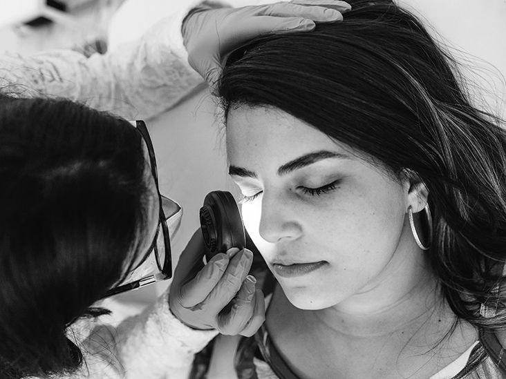
Diagnosis starts with suspicion. A dermatologist examines the lesion, asks how it has changed, and often looks at it with a dermatoscope, a handheld magnifying tool with light that helps reveal structures not obvious to the naked eye. Dermoscopy can improve the odds of spotting a problem, but it is still not the final answer.
The only way to know for sure is a biopsy. In many cases, the doctor removes all or part of the suspicious lesion and sends it to a lab. Pathologists then determine whether it is melanoma and, if so, how thick it is, whether ulceration is present, and other features that help guide staging and treatment.
If melanoma is confirmed, the next steps may include wider excision of the area, imaging in selected cases, and sometimes sentinel lymph node biopsy depending on the thickness and other tumor features. In plain English: first confirm what it is, then find out how far it has gone, if anywhere.
Prognosis: What It Really Depends On
When people ask about prognosis, they usually want a simple number. Melanoma, unhelpfully, prefers nuance.
The most important factors in prognosis include:
- Stage at diagnosis
- Tumor thickness (often called Breslow depth)
- Whether the lesion is ulcerated
- Whether lymph nodes are involved
- Whether the cancer has spread to distant organs
- How quickly the lesion was identified and treated
Studies comparing amelanotic melanoma with pigmented melanoma suggest that the amelanotic form often has worse outcomes at diagnosis largely because it is found later and is more advanced or thicker by the time it is recognized. That is an important distinction. The problem is not just the biology of the tumor. It is also the delay caused by the disguise.
What survival data tells us
For melanoma overall in the United States, the outlook is strongly tied to stage. When melanoma is localized to the original site, five-year relative survival is excellent. Once it has spread regionally or distantly, survival drops. That does not mean late-stage cases are hopeless. It means stage changes the game.
Modern treatment has improved the outlook for advanced melanoma more than many people realize. Immunotherapy and targeted therapy have transformed care for some patients with stage IV disease, and long-term survival for advanced melanoma is better than it used to be. Still, the best prognosis usually comes from catching melanoma before it has spread.
So if you want the plain-language version, here it is: amelanotic melanoma can have a good prognosis when found early, and a tougher prognosis when found late. That may sound obvious, but with this subtype, that simple truth matters more than ever.
Treatment Basics
Treatment depends on stage. Early lesions are often treated with surgery to remove the melanoma along with a margin of normal skin. If the melanoma is thicker or shows high-risk features, doctors may recommend additional procedures to check nearby lymph nodes.
More advanced cases may be treated with:
- Immunotherapy
- Targeted therapy for tumors with certain gene changes
- Radiation in selected situations
- Clinical trials
Follow-up matters too. People who have had one melanoma are at increased risk for another. That means regular skin exams, sun protection, and learning your own skin pattern are not optional extras. They are part of the long game.
How to Check Your Skin Without Turning Into a Full-Time Detective
You do not need a medical degree or a magnifying headset worthy of a crime show. A monthly self-exam in good lighting is a smart habit. Use a full-length mirror and a hand mirror. Check your face, ears, scalp, back, buttocks, legs, soles, palms, and nails. Ask for help with hard-to-see areas if needed.
Pay attention to any spot that:
- Looks different from the others
- Changes over time
- Itches, hurts, or bleeds
- Does not heal
- Seems new and oddly persistent
Take a photo if you are unsure. Not to diagnose yourself, but to compare whether it changes. Skin has a memory problem. Your camera does not.
Experiences People Commonly Have With Amelanotic Melanoma
One of the most frustrating experiences people describe with amelanotic melanoma is that it simply does not look scary at first. There may be no dramatic dark color, no obvious “bad mole” vibe, and no instant alarm bell. Instead, the lesion can seem like a minor annoyance: a pink bump after shaving, a sore spot on the scalp, a flaky patch near the ankle, or a little area that looks irritated from friction. Because it seems ordinary, people often watch it for a while. Then they watch it some more. Then they make a mental note to deal with it after work gets less busy, after the holidays, after the next vacation, after literally anything else.
Another common experience is confusion. A person may treat the spot like acne, dry skin, or a wound. They may apply moisturizer, antibiotic ointment, or over-the-counter cream and feel encouraged when it seems a little better for a few days. Then it bleeds again, crusts again, or keeps growing. That on-and-off behavior can be misleading. It creates false reassurance, and false reassurance is one of melanoma’s favorite tricks.
People also describe feeling surprised when the lesion is biopsied. Many expect the result to be something minor because the spot never matched the classic image of melanoma. That emotional whiplash can be intense. One week it is “probably nothing,” and the next week they are hearing words like margins, thickness, stage, and lymph node. The jump from ordinary skin issue to cancer discussion can feel surreal.
For caregivers and family members, the experience is often a mix of guilt and vigilance. They may think, “How did we not notice this sooner?” The honest answer is that amelanotic melanoma is easy to underestimate. It is not a failure of character to miss a lesion that looks like a harmless spot. The important part is what happens next: getting a diagnosis, getting treatment, and sticking with follow-up.
After treatment, many people become more tuned in to their skin than ever before. That can be empowering, but it can also make every new bump feel suspicious. The challenge is finding a balance between healthy awareness and constant panic. A good dermatologist can help patients build that balance. So can photos, body maps, and a routine self-exam schedule. Structure helps keep worry from running the whole show.
There is also a practical side to the experience. Lesions on the scalp may be found only during a haircut or by a partner. Lesions on the foot may be mistaken for a blister or bruise from shoes. Nail lesions may be dismissed as trauma. In each case, the story is similar: the melanoma hides in plain sight by looking like something common. That is why awareness matters so much. Not because every odd pink spot is cancer, but because some of them are, and amelanotic melanoma benefits when people do not know it exists.
Many survivors and clinicians come back to the same lesson: trust change. A spot that changes, persists, bleeds, or feels different deserves more respect than a casual shrug. With amelanotic melanoma, that small decision to get a strange lesion checked can make a very big difference.
Final Takeaway
Amelanotic melanoma is uncommon, but it earns attention because it hides behind a less alarming appearance. It may be pink, red, white, skin-colored, or almost clear. It may look like a pimple, scar, cyst, or sore. That disguise is exactly why it can be diagnosed later than classic melanoma.
The good news is that early detection still changes everything. A spot that evolves, bleeds, does not heal, or just looks different from your other spots is worth a professional evaluation. A biopsy, not guesswork, gives the answer. And when melanoma is found early, the outlook is dramatically better.
So no, you do not need to panic over every pink bump. But you also should not let a persistently weird lesion keep freeloading on your skin without an explanation.