Note: This article is for educational purposes only. It explains lower leg anatomy in clear, reader-friendly language and should not replace medical advice, diagnosis, or treatment from a qualified healthcare professional.
The lower leg may look like a simple stretch of shin, calf, ankle, and the occasional mystery bruise from a coffee table you swear moved on its own. But anatomically, it is a remarkably organized structure. It carries body weight, absorbs shock, helps you walk, run, jump, balance, climb stairs, stand on tiptoe, and perform that awkward little jog when someone holds the elevator door.
In strict anatomical language, the “leg” is the region between the knee and the ankle. What many people casually call the “whole leg” is actually the lower limb, which includes the hip, thigh, knee, leg, ankle, and foot. The lower leg itself contains two bones, several muscle compartments, major nerves, arteries, veins, tendons, ligaments, fascia, and skin. Each part has a job, and when all parts cooperate, movement feels effortless. When one part complains, suddenly walking to the fridge becomes a dramatic athletic event.
Lower Leg Anatomy at a Glance
The lower leg is built around the tibia and fibula. The tibia is the larger, weight-bearing bone on the inner side of the lower leg, while the fibula is the slimmer bone on the outer side. These bones connect the knee to the ankle and provide attachment points for muscles, tendons, and ligaments.
A helpful way to understand lower leg anatomy is to think of it as a well-designed engineering project. The bones form the frame, muscles create movement, tendons transfer force, nerves deliver instructions, arteries bring oxygen-rich blood, veins return blood to the heart, and fascia wraps everything into neat compartments. It is less like a random pile of body parts and more like a high-performance suspension system wearing socks.
The Bones of the Lower Leg
The Tibia: The Shin Bone and Main Support Beam
The tibia, commonly called the shin bone, is the larger and stronger of the two lower leg bones. It sits on the medial side, meaning closer to the midline of the body. You can feel its front edge just under the skin along your shin. That is why bumping your shin on a bed frame feels like receiving a personal message from your skeleton.
The tibia carries most of the body’s weight as it passes from the thigh into the foot. At its upper end, the tibia helps form the knee joint with the femur, or thigh bone. At its lower end, it helps form the ankle joint with the fibula and talus. The inner ankle bump, called the medial malleolus, is part of the tibia.
The tibia also provides attachment sites for important muscles and ligaments. Because it plays such a central role in standing and walking, tibial injuries can affect nearly every daily movement, from climbing stairs to getting out of a chair.
The Fibula: The Slim Stabilizer
The fibula is the thinner bone on the outer side of the lower leg. It does not carry as much body weight as the tibia, but do not mistake “smaller” for “unimportant.” The fibula helps stabilize the ankle, supports muscle attachments, and forms the outer ankle bump, known as the lateral malleolus.
The fibula works with the tibia through connective tissue and joints. Between the two bones is the interosseous membrane, a strong sheet of tissue that helps hold them together and provides additional attachment space for muscles. This membrane also helps distribute forces between the tibia and fibula during movement.
The Ankle Connection: Where the Lower Leg Meets the Foot
The ankle joint is formed mainly by the tibia, fibula, and talus. The lower ends of the tibia and fibula create a socket-like structure called the ankle mortise, which holds the talus. This design allows the foot to move up and down in motions called dorsiflexion and plantar flexion.
Dorsiflexion happens when you lift your toes toward your shin. Plantar flexion happens when you point your toes downward, like pressing a gas pedal or standing on tiptoe. These movements may seem simple, but they require coordinated work from bones, muscles, tendons, ligaments, and nerves.
Muscle Compartments of the Lower Leg
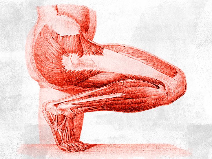
Lower leg muscles are grouped into compartments. These compartments are separated by fascia, a strong connective tissue that wraps around muscles and keeps them organized. Most anatomy references describe the lower leg in four functional compartments: anterior, lateral, superficial posterior, and deep posterior.
Anterior Compartment: The Toe-Lifting Team
The anterior compartment sits at the front of the lower leg. Its main job is dorsiflexion, which lifts the foot upward. This compartment includes muscles such as the tibialis anterior, extensor hallucis longus, extensor digitorum longus, and fibularis tertius.
The tibialis anterior is especially important for walking because it helps lift the foot so the toes clear the ground. Without it, the foot may slap down or drag during each step. The extensor muscles also help lift the big toe and the other toes. These muscles are small compared with the calf, but they are essential for smooth, controlled walking.
Lateral Compartment: The Side-to-Side Stabilizers
The lateral compartment lies along the outer side of the lower leg. It contains the fibularis longus and fibularis brevis, also called the peroneus longus and peroneus brevis. These muscles help evert the foot, meaning they turn the sole slightly outward. They also assist with plantar flexion and provide ankle stability.
If you step on uneven ground, the lateral compartment helps your ankle adjust quickly. It is one of the reasons you can walk across grass, gravel, or a slightly suspicious sidewalk without instantly toppling over like a folding chair.
Superficial Posterior Compartment: The Powerful Calf
The posterior compartment is located at the back of the lower leg. Its superficial layer contains the big, visible calf muscles: the gastrocnemius and soleus. Together, these muscles are often called the triceps surae. They join into the Achilles tendon, which attaches to the heel bone, or calcaneus.
The gastrocnemius crosses both the knee and ankle, helping bend the knee and point the foot downward. The soleus lies deeper and is especially important for standing and walking. While the gastrocnemius gets more attention because it shapes the calf, the soleus is the quiet workhorse that helps keep you upright for long periods.
Deep Posterior Compartment: The Hidden Control Center
The deep posterior compartment includes muscles such as the tibialis posterior, flexor hallucis longus, flexor digitorum longus, and popliteus. These muscles support the arch of the foot, flex the toes, assist with plantar flexion, and help control ankle and foot position.
The tibialis posterior is particularly important for arch support and foot stability. When this muscle or its tendon becomes weak or irritated, the arch can flatten and walking may become painful. The flexor hallucis longus helps flex the big toe, which is a surprisingly important action during push-off in walking and running.
Tendons and Ligaments in the Lower Leg
Tendons connect muscles to bones. In the lower leg, tendons act like strong cables that transfer muscle force to the foot and ankle. The Achilles tendon is the most famous example. It allows the calf muscles to lift the heel, push the body forward, and power movements such as jumping, sprinting, and climbing.
Ligaments connect bones to bones. Around the ankle, ligaments help hold the tibia, fibula, and talus in proper alignment. They protect the joint from excessive motion. When an ankle twists too far, these ligaments can stretch or tear, causing a sprain. That is why ankle sprains are so common in sports, hiking, and heroic attempts to carry all grocery bags in one trip.
Nerves of the Lower Leg
Nerves are the communication lines of the lower leg. They carry signals from the brain and spinal cord to the muscles and return sensory information from the skin, joints, and tissues.
The Tibial Nerve
The tibial nerve travels through the back of the leg and supplies many of the posterior compartment muscles. It helps control plantar flexion, toe flexion, and important foot muscles. It also contributes to sensation in parts of the lower leg and foot.
The Common Fibular Nerve
The common fibular nerve, also called the common peroneal nerve, travels around the outside of the knee near the fibular head. Because it runs close to the surface in this area, it can be vulnerable to injury or compression. It divides into the deep fibular nerve and superficial fibular nerve.
The deep fibular nerve supplies the anterior compartment and helps lift the foot and toes. The superficial fibular nerve supplies the lateral compartment and helps with foot eversion. When these nerves are affected, symptoms may include weakness, tingling, numbness, or changes in walking pattern.
Sensory Nerves and Skin Feeling
Several smaller sensory nerves help the lower leg detect touch, pressure, temperature, and pain. The saphenous nerve contributes sensation along the inner lower leg, while the sural nerve supplies parts of the outer and back lower leg and foot. This sensory map matters clinically because the location of numbness or tingling can help healthcare professionals identify which nerve may be irritated.
Blood Vessels of the Lower Leg
The lower leg needs a steady blood supply to keep muscles, nerves, bones, and skin healthy. The main arterial pathway begins behind the knee, where the popliteal artery branches into vessels that supply the lower leg and foot.
Major Arteries
The anterior tibial artery supplies the front of the lower leg and continues into the foot as the dorsalis pedis artery. The posterior tibial artery supplies the back and bottom-side structures of the leg and foot. The fibular artery, also called the peroneal artery, supplies deeper and lateral structures.
These arteries deliver oxygen and nutrients during rest and activity. When you run, climb, or jump, the muscles demand more oxygen, and blood flow increases to meet the need. If circulation is reduced, symptoms may include cramping, fatigue, coldness, or pain during activity.
Veins and Blood Return
Veins return blood from the lower leg to the heart. Deep veins generally travel with the arteries, while superficial veins lie closer to the skin. The lower leg also depends on the “calf muscle pump.” Each time the calf muscles contract, they help push blood upward against gravity. This is one reason walking and ankle movement can support circulation, especially during long periods of sitting.
Fascia and Compartments: The Body’s Internal Organizers
Fascia is connective tissue that surrounds and separates muscles. In the lower leg, fascia forms compartments that keep muscle groups in place. This organization is useful, but it also explains a serious condition called compartment syndrome.
Compartment syndrome occurs when pressure rises inside a closed muscle compartment. Because fascia does not stretch much, swelling can compress blood vessels and nerves. Acute compartment syndrome is a medical emergency and may happen after fractures, crush injuries, or severe trauma. Symptoms can include intense pain, tightness, numbness, weakness, and pain with stretching the affected muscles.
Common Lower Leg Conditions Linked to Anatomy
Shin Splints
Shin splints, often called medial tibial stress syndrome, usually cause pain along the inner edge of the tibia. They are common in runners, dancers, military trainees, and anyone who suddenly increases training intensity. The pain is linked to stress on muscles, tendons, and bone tissue around the tibia.
Calf Strain
A calf strain involves overstretching or tearing fibers in the gastrocnemius or soleus. It may happen during sprinting, jumping, or sudden acceleration. A person may feel a sharp pull or popping sensation, followed by pain, swelling, or difficulty pushing off the foot.
Achilles Tendon Problems
The Achilles tendon handles tremendous force. Overuse, tight calf muscles, poor training progression, or sudden explosive movement can irritate or injure it. Achilles tendinopathy often causes pain and stiffness near the back of the heel, especially after rest or during activity.
Fractures and Stress Injuries
The tibia and fibula can fracture from trauma, falls, sports injuries, or repetitive stress. A tibial stress fracture may begin as activity-related pain that worsens over time. Because the tibia bears significant weight, persistent shin pain should not be ignored.
How the Lower Leg Works During Walking
Walking is a beautiful example of lower leg anatomy in action. When your heel strikes the ground, the anterior compartment helps control the foot so it does not slap down. As your body moves forward, the tibia and fibula transfer load toward the ankle and foot. The calf muscles then contract, the Achilles tendon tightens, and the foot pushes off the ground.
During this process, nerves coordinate timing, arteries deliver oxygen, veins return blood, ligaments stabilize joints, and fascia keeps muscle groups organized. All of that happens with each step. The lower leg is basically doing a tiny engineering miracle while you are thinking about lunch.
Everyday Experiences That Make Lower Leg Anatomy Easier to Understand
One of the best ways to understand lower leg anatomy is to notice it during normal life. You do not need a cadaver lab, a medical textbook, or a dramatic sports injury montage. You can learn a lot by walking around the block, climbing stairs, standing in line, or trying to balance on one foot while putting on a shoe.
For example, think about walking uphill. Your calf muscles work harder because they must lift the heel and push the body upward. The gastrocnemius and soleus contract again and again, while the Achilles tendon transfers that force to the heel. After a steep hill, your calves may feel like they have submitted a formal complaint. That sensation is a practical reminder of how powerful the posterior compartment is.
Now imagine walking downhill. The front of the lower leg becomes more noticeable. The tibialis anterior helps control the foot as it lowers to the ground. Without that controlled action, every step would land with a slap. If you have ever felt soreness along the front of your shins after a long downhill hike, you have met your anterior compartment in a very memorable way.
Standing for a long time tells another story. The soleus muscle helps maintain posture and supports venous return through the calf pump. When you shift your weight from one foot to the other, rise slightly onto your toes, or flex your ankles while waiting in a line, you are not just fidgeting. You are helping muscles and veins move blood upward. Your body is quietly saying, “Please do not make me stand like a statue.”
Running highlights the lower leg’s shock-absorbing role. Each stride sends force through the foot, ankle, tibia, and surrounding muscles. The tibia handles load, the fibula helps stabilize the ankle, and the calf muscles drive push-off. If training increases too quickly, tissues may not adapt fast enough. That is why gradual progression matters. Anatomy likes ambition, but it prefers ambition with warm-ups and sensible shoes.
Even footwear reveals lower leg mechanics. Shoes with very high heels keep the ankle in plantar flexion, shortening the calf muscles during wear. Flat, unsupportive shoes may increase strain in people who need more arch or ankle support. The lower leg constantly adapts to what is under the foot, whether that is a running shoe, work boot, sandal, or the questionable house slipper you refuse to retire.
Balance is another everyday anatomy lesson. Stand on one foot and you will feel tiny corrections around the ankle. The lateral compartment helps prevent excessive rolling outward, while deep posterior muscles help control the arch and foot position. Nerves send rapid updates about pressure, stretch, and position. Your brain processes all of it quickly, often before you consciously realize you are wobbling.
Finally, lower leg anatomy shows up when something feels “off.” A tight calf, sore shin, numb outer foot, swollen ankle, or aching Achilles tendon can each point to a different structure. Understanding the basics helps you describe symptoms more clearly. Instead of saying “my leg thing hurts,” you might recognize whether the discomfort is near the shin bone, calf muscle, ankle tendon, or outside of the fibula. That vocabulary can make conversations with trainers, physical therapists, and healthcare professionals much more useful.
Conclusion
The anatomy of the lower leg is a compact masterpiece of support, motion, balance, and communication. The tibia carries weight, the fibula stabilizes the ankle, muscles move the foot and toes, tendons transfer force, ligaments protect joints, nerves coordinate action, and blood vessels keep tissues supplied. Whether you are walking to work, running a race, dancing in the kitchen, or simply standing in line for coffee, your lower leg is doing far more than it gets credit for.
Understanding lower leg anatomy can also help you appreciate common problems such as shin splints, calf strains, Achilles tendon pain, ankle sprains, nerve irritation, and circulation issues. The lower leg may not be flashy, but it is essential. Treat it well with smart movement, gradual training, supportive footwear, and attention to persistent pain. After all, these hardworking structures carry you through the world every day, and they rarely ask for more than a little stretching and fewer surprise encounters with furniture.